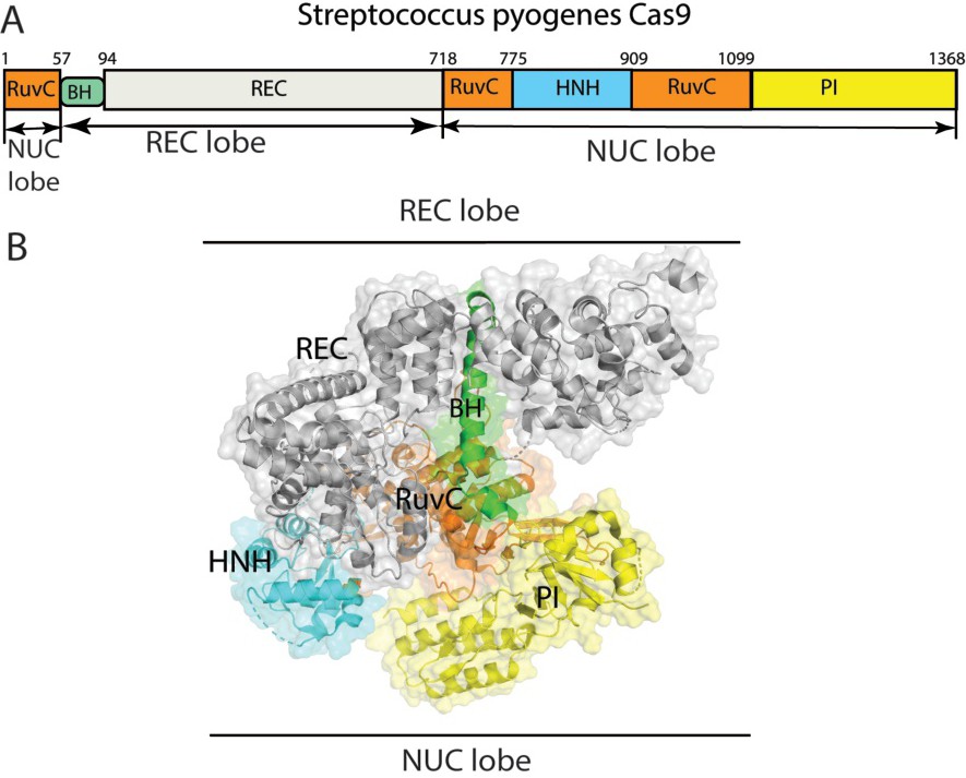
Fig. 1. A. Schematic illustration of domain organization for the type II spCas9. B. Cartoon-surface representations of the three-dimensional structure of spCas9 ID 4CMP from the Protein Data Bank (PDB) database. Structural image was prepared with the PyMOL Molecular Graphics System, Version 2.0 Schrödinger, LLC and Adobe Illustrator.